scroll down for posts
I think it's very important to have a feedback loop, where you're constantly thinking about what you've done and how you could be doing it better. I think that's the single best piece of advice: constantly think about how you could be doing things better and questioning yourself.
Elon Musk
But what has he achieved?
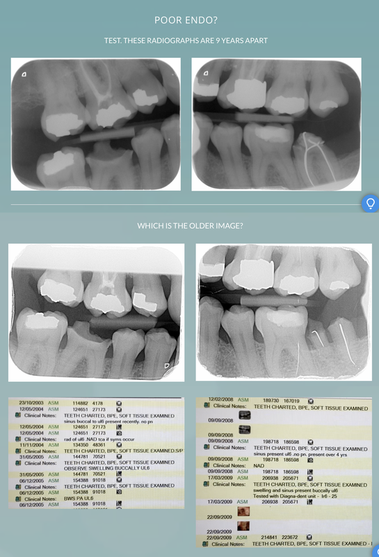
I made this for my FDs and yours, please support them through Peer review groups. Some blogs may be a starting point for discussion. We can improve their working environment, reduce their stress, and enjoy their dentistry as I have, long enough to look after my teeth.
 RSS Feed